.avif)
Digital X-rays in Longmont, CO | Martin Family Orthodontics
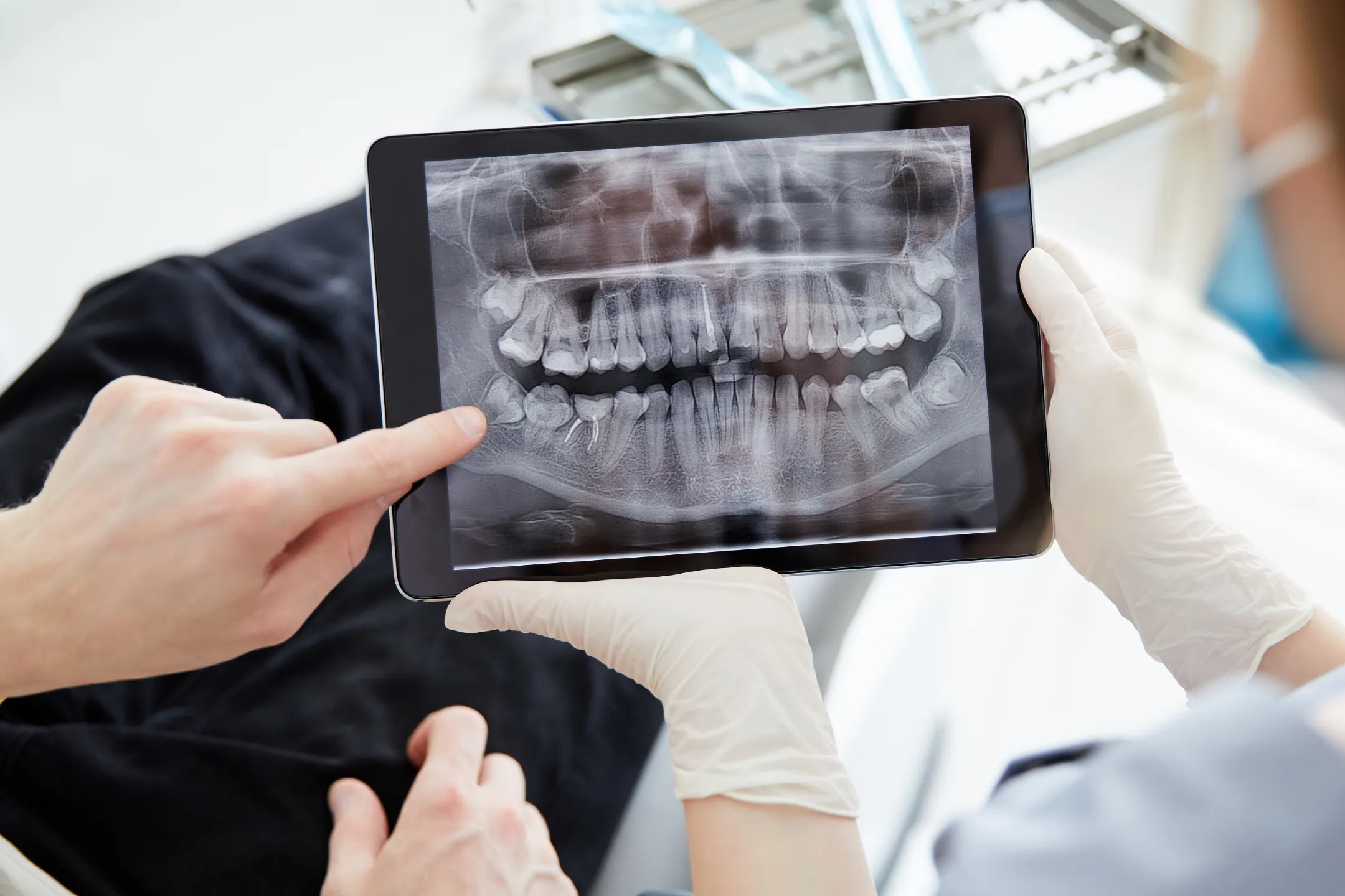
Digital X-rays at Martin Family Orthodontics in Longmont, CO replace traditional film X-rays with a modern imaging system that uses dramatically less radiation, produces clearer images, and lets us review and share results instantly. For orthodontic patients of any age, digital radiography is the safer, faster, more informative option.
What Are Digital X-rays?
Digital X-rays use an electronic sensor in place of traditional photographic film. When an X-ray is taken, the sensor captures the image digitally and displays it on a computer screen within seconds. Compared to film, the radiation dose is significantly lower, the resolution is higher, and the images can be magnified, contrast-adjusted, and annotated for clearer diagnosis.
Why Digital X-rays Matter for Orthodontic Treatment
Orthodontic treatment depends on accurate imaging. Before we move teeth, we need to know exactly where they are, what their roots look like, and what surrounding structures we are working around. Digital X-rays give Dr. Laurel Martin detailed information at every stage of care, including:
- Initial diagnosis. Identifying tooth positions, root angulation, and developmental issues.
- Pre-treatment planning. Confirming the position of impacted or unerupted teeth, especially in younger patients.
- Progress monitoring. Verifying that teeth are tracking correctly during treatment.
- Post-treatment evaluation. Confirming root health and final alignment.
For more complex cases, we may supplement standard digital X-rays with 3D CT scanning or panoramic X-rays to get the complete picture.
Benefits of Digital X-rays
Less radiation than film. Digital sensors require dramatically less radiation than older film systems, often 50 to 80 percent less. The exposure for a single digital X-ray is comparable to the natural background radiation you receive in a couple of days.
Instant results. Images appear on the screen within seconds. There is no waiting for film to develop, no chemical processing, and no chance the image needs to be retaken because of poor development.
Clearer diagnostic detail. Digital images can be magnified, sharpened, and adjusted for contrast. Small details such as early decay, root resorption, or hairline root fractures are easier to identify.
Easy sharing with other providers. Digital files can be sent to your general dentist, an oral surgeon, or any other provider involved in your care without the delays of mailing physical film.
Environmentally cleaner. No chemical developing fluids and no disposable film.
Types of Digital X-rays We Use
Bitewing X-rays. Capture the crowns of upper and lower teeth in a single image. Useful for assessing tooth position, interproximal contact, and signs of decay.
Periapical X-rays. Capture a single tooth or a small group of teeth from crown to root tip. These are the go-to images for evaluating root position and length during orthodontic care.
Cephalometric X-rays. Side-profile views of the head used to evaluate the relationship between the upper and lower jaws, tooth angulation, and skeletal proportions. Essential for treatment planning and for tracking changes over time.
Panoramic X-rays. Single image showing all teeth, jaws, sinuses, and TMJ structures. Covered in more detail on our panoramic X-rays page.
Are Digital X-rays Safe?
Yes. Modern digital radiography uses very low radiation doses, and we follow ALARA principles (As Low As Reasonably Achievable) for every imaging decision. We only take X-rays when the diagnostic information is necessary for your care, and we use lead shields and thyroid collars to protect surrounding tissue.
Children and pregnant patients receive additional consideration. We can defer non-urgent imaging when appropriate, and we will discuss any concerns directly with you.
How Often Do You Need X-rays During Orthodontic Treatment?
The frequency depends on your case. Most patients have a complete set at the start of treatment, periodic check images during active treatment if clinically needed, and a final set near completion. Routine monitoring usually does not require frequent imaging.
We do not over-image. Every X-ray request is tied to a specific clinical question. If the answer to a question does not require imaging, we do not take the picture.
Frequently Asked Questions About Digital X-rays
Heading
Lorem ipsum dolor sit amet, consectetur adipiscing elit, sed do eiusmod tempor incididunt ut labore et dolore magna aliqua. Ut enim ad minim veniam, quis nostrud exercitation ullamco laboris nisi ut aliquip ex ea commodo consequat. Duis aute irure dolor in reprehenderit in voluptate velit esse cillum dolore eu fugiat nulla pariatur.
How long does it take to get a digital X-ray?
The exposure itself takes a fraction of a second. The image appears on screen within a few seconds.
Will I be charged extra for X-rays?
Diagnostic imaging is typically included in the cost of comprehensive treatment or billed as part of standard exam codes. We will discuss costs and coverage during onboarding.
Are digital X-rays covered by insurance?
Most dental insurance plans cover diagnostic imaging at standard intervals. Our team will verify your specific benefits.
Can I see my X-rays?
Yes. We will walk you through your images at your consultation and explain what we are seeing.
Can my general dentist use these X-rays?
Yes. With your consent, we share digital imaging with other providers involved in your care.
Schedule a Consultation
If you are starting orthodontic treatment or want a second opinion on a case in progress, a consultation at Martin Family Orthodontics gives you access to modern diagnostic imaging and the experienced clinical judgment to interpret it well.